MIT: Ultrasound Adhesives for Imaging Organs
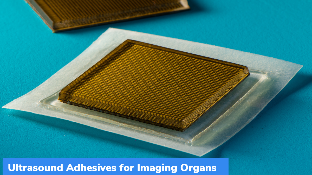
Researchers at Massachusetts Institute of Technology (MIT) has developed a postage stamp-sized device. This device can create live, high-resolution images. This device can be affixed to the skin and is capable of transmitting images continuously for 48 hours.
Important facts about Ultrasound Adhesives:
- Using Ultrasound Adhesives, one can see internal organs with few patches on the body.
- The sticker is around 3 to 4 inches across and 1/10-inch thick.
- It will substitute the bulky, specialized ultrasound equipment used at hospitals and doctor’s clinics. On the equipment, technicians apply a gel on skin and then use a wand or probe to send sound waves into the body.
- Sound waves reflect back high-resolution images of blood vessels and organs like lungs, heart, and stomach.
- Some hospitals have affixed the equipment to robotic arms, to provide imaging for extended periods.
- For now, the stickers are needed to be connected to instruments. Researchers are working to find a way to operate them wirelessly.
- The current design of Ultrasound Adhesives could eliminate the requirement for technician to hold it in place for a long time.
During the study, these patches adhered to the skin well. It enabled the researchers to capture images even if the patient was sitting, standing, jogging and biking. Wearable ultrasound imaging equipment have huge potential in clinical diagnosis. However, resolution and imaging duration of newly developed ultrasound patches is currently low, and thus cannot image deep organs.
Month: Current Affairs - August, 2022
Category: Science & Technology Current Affairs


